
Bio
Dr. Mohammad Abu Sayem Karal was born in Sadarpur under Faridpur district, Bangladesh on 10th December 1980. He obtained his B.Sc.(Honours) in 2001 (examination held in 2004) and M. S. in 2002 (examination held in 2005) in Physics from the Department of Physics, University of Dhaka, Bangladesh. He received his M.Phil degree in Physics from the Department of Physics, Bangladesh University of Engineering and Technology (BUET), Dhaka in 2011. He joined the Department of Physics, BUET in 18th February, 2008 as a Lecturer. He became Assistant Professor on 28th December, 2011, Associate Professor on 12th March, 2018 and Professor on 20th December, 2020. He obtained Ph. D. degree in the field of Membrane Biophysics from the Department of Bioscience, Shizuoka University, Japan in September 2015. He received Monbukagakusho (MEXT) scholarship for his Ph. D study. His present research interests are Membrane Biophysics (Experimental and Simulation) including Protein-Lipid interactions, Nanobiotechnology, Focused Impedance Method (FIM), Correlation between MRI and Nerve Conduction Velocity (NCV), and Radiation Biophysics. He secured BCS cadre (First Position in Physics in 26th BCS). His spouse is a BCS cadre officer under the Ministry of Education. He has two daughters.
Education & Training
-
Ph.D 2015
Biophysics
Department of Bioscience, Graduate School of Science and Technology, Shizuoka University, Japan.
-
M.Phil. 2011
Material Science
Department of Physics, Bangladesh University of Engineering and Technology (BUET)
-
MS 2005
Biomedical Physics
Department of Physics, University of Dhaka
-
B.Sc. (Hons.) 2003
Physics
Department of Physics, University of Dhaka
Honors, Awards and Grants
Publications
Filter by type:
Sort by year:
Effects of cholesterol on the size distribution and bending modulus of lipid vesicles
Journal Paper PLoS ONE 17(1): e0263119 (2022)
Abstract
The influence of cholesterol fraction in the membranes of giant unilamellar vesicles (GUVs) on their size distributions and bending moduli has been investigated. The membranes of GUVs were synthesized by a mixture of two elements: electrically neutral lipid 1, 2-dioleoyl-sn-glycero-3-phosphocholine (DOPC) and cholesterol and also a mixture of three elements: electrically charged lipid 1,2-dioleoyl-sn-glycero-3-phospho-(1′-rac-glycerol) (DOPG), DOPC and cholesterol. The size distributions of GUVs have been presented by a set of histograms. The classical lognormal distribution is well fitted to the histograms, from where the average size of vesicle is obtained. The increase of cholesterol content in the membranes of GUVs increases the average size of vesicles in the population. Using the framework of Helmholtz free energy of the system, the theory developed by us is extended to explain the experimental results. The theory determines the influence of cholesterol on the bending modulus of membranes from the fitting of the proper histograms. The increase of cholesterol in GUVs increases both the average size of vesicles in population and the bending modulus of membranes.
Quantification of pulsed electric field for the rupture of giant vesicles with various surface charges, cholesterols and osmotic pressures
Journal Paper PLoS ONE 17(1): e0262555 (2022)
Abstract
Electropermeabilization is a promising phenomenon that occurs when pulsed electric field with high frequency is applied to cells/vesicles. We quantify the required values of pulsed electric fields for the rupture of cell-sized giant unilamellar vesicles (GUVs) which are prepared under various surface charges, cholesterol contents and osmotic pressures. The probability of rupture and the average time of rupture are evaluated under these conditions. The electric field changes from 500 to 410 Vcm-1 by varying the anionic lipid mole fraction from 0 to 0.60 for getting the maximum probability of rupture (i.e., 1.0). In contrast, the same probability of rupture is obtained for changing the electric field from 410 to 630 Vcm-1 by varying the cholesterol mole fraction in the membranes from 0 to 0.40. These results suggest that the required electric field for the rupture decreases with the increase of surface charge density but increases with the increase of cholesterol. We also quantify the electric field for the rupture of GUVs containing anionic mole fraction of 0.40 under various osmotic pressures. In the absence of osmotic pressure, the electric field for the rupture is obtained 430 Vcm-1, whereas the field is 300 Vcm-1 in the presence of 17 mOsmL-1, indicating the instability of GUVs at higher osmotic pressures. These investigations open an avenue of possibilities for finding the electric field dependent rupture of cell-like vesicles along with the insight of biophysical and biochemical processes.
Recent development on the kinetics of rupture of giant vesicles under constant tension
Journal Paper RSC Adv, 11(47), 29598-29619 (2021)
Abstract
External tension in membranes plays a vital role in numerous physiological and physicochemical phenomena. In this review, recent developments in the constant electric- and mechanical-tension-induced rupture of giant unilamellar vesicles (GUVs) are considered. We summarize the results relating to the kinetics of GUV rupture as a function of membrane surface charge, ions in the bathing solution, lipid composition, cholesterol content in the membrane, and osmotic pressure. The mechanical stability and line tension of the membrane under these conditions are discussed. The membrane tension due to osmotic pressure and the critical tension of rupture for various membrane compositions are also discussed. The results and their analysis provide a biophysical description of the kinetics of rupture, along with insight into biological processes. Future directions and possible developments in this research area are included.
A new purification technique to obtain specific size distribution of giant lipid vesicles using dual filtration
Journal Paper PLoS ONE, 16(7): e0254930 (2021)
Abstract
A new purification technique is developed for obtaining distribution of giant unilamellar vesicles (GUVs) within a specific range of sizes using dual filtration. The GUVs were prepared using well known natural swelling method. For filtration, different combinations of polycarbonate membranes were implemented in filter holders. In our experiment, the combinations of membranes were selected with corresponding pore sizes–(i) 12 and 10 μm, (ii) 12 and 8 μm, and (iii) 10 and 8 μm. By these filtration arrangements, obtained GUVs size distribution were in the ranges of 6−26 μm, 5–38 μm and 5–30 μm, respectively. In comparison, the size distribution range was much higher for single filtration technique, for example, 6−59 μm GUVs found for a membrane with 12 μm pores. Using this technique, the water-soluble fluorescent probe, calcein, can be removed from the suspension of GUVs successfully. The size distributions were analyzed with lognormal distribution. The skewness became smaller (narrow size distribution) when a dual filtration was used instead of single filtration. The mode of the size distribution obtained in dual filtration was also smaller to that of single filtration. By continuing this process of purification for a second time, the GUVs size distribution became even narrower. After using an extra filtration with dual filtration, two different size distributions of GUVs were obtained at a time. This experimental observation suggests that different size specific distributions of GUVs can be obtained easily, even if GUVs are prepared by different other methods.
Effects of osmotic pressure on the irreversible electroporation in giant lipid vesicles
Journal Paper PLoS ONE 16(5): e0251690, 2021.
Abstract
Irreversible electroporation (IRE) is a nonthermal tumor/cell ablation technique in which a series of high-voltage short pulses are used. As a new approach, we aimed to investigate the rupture of giant unilamellar vesicles (GUVs) using the IRE technique under different osmotic pressures (Π), and estimated the membrane tension due to Π. Two categories of GUVs were used in this study. One was prepared with a mixture of dioleoylphosphatidylglycerol (DOPG), dioleoylphosphatidylcholine (DOPC) and cholesterol (chol) for obtaining more biological relevance while other with a mixture of DOPG and DOPC, with specific molar ratios. We determined the rate constant (kp) of rupture of DOPG/DOPC/chol (46/39/15)-GUVs and DOPG/DOPC (40/60)-GUVs induced by constant electric tension (σc) under different Π. The σc dependent kp values were fitted with a theoretical equation, and the corresponding membrane tension (σoseq) at swelling equilibrium under Π was estimated. The estimated membrane tension agreed well with the theoretical calculation within the experimental error. Interestingly, the values of σoseq were almost same for both types of synthesized GUVs under same osmotic pressure. We also examined the sucrose leakage, due to large osmotic pressure-induced pore formation, from the inside of DOPG/DOPC/chol(46/39/15)-GUVs. The estimated membrane tension due to large Π at which sucrose leaked out was very similar to the electric tension at which GUVs were ruptured without Π. We explained the σc and Π induced pore formation in the lipid membranes of GUVs.
Analysis of purification of charged giant vesicles in a buffer using their size distribution
Journal Paper Eur. Phys. J. E 44, 62, 2021.
Abstract
We have analyzed the purification of charged giant unilamellar vesicles (GUVs) prepared in a buffer containing various concentrations of salt using their size distribution. The membranes of GUVs were synthesized by a mixture of dioleoylphosphocholine (DOPC) and dioleoylphosphatidylglycerol (DOPG) lipids. The DOPG mole fractions (X) in the membranes of GUVs were 0.10, 0.25, 0.40, 0.55, 0.70, 0.90 in a physiological buffer containing 162 mM salt. In addition, for a fixed value of X the concentrations of salt (C) in the buffer were 12, 62, 112, 162, 212, 312, 362 mM. The size distribution histograms of experimentally investigated unpurified and purified GUVs were fitted with the lognormal distribution and obtained the multiplication factor γ for mean (μ) and η for standard deviation (σ) of the lognormal distribution. The key parameters γ and η were responsible for changing the average size and size distribution of unpurified GUVs to purified ones. The theoretically fitting equation of experimentally obtained X- and C-dependent values of γ and η provided the calibration equation for estimating the average size of purified GUVs theoretically for any values of X and C. The estimated size of purified GUVs increased with the increase in electrostatic effect (i.e., increase in vesicle surface charge density or decrease in salt concentration in buffer). The estimated size of purified GUVs varied with X and C, which supported the previous report qualitatively. These investigations might be helpful in the field of cell/chemical biology for understanding the process of purification of vesicles/cells investigated by any other techniques.
An investigation into the critical tension of electroporation in anionic lipid vesicles
Journal Paper Eur Biophys J 50, 99–106, 2021.
Abstract
Irreversible electroporation (IRE) is a technique for the disruption of localized cells or vesicles by a series of short and high−frequency electric pulses which has been used for tissue ablation and treatment in certain diseases. It is well reported that IRE induces lateral tension in the membranes of giant unilamellar vesicles (GUVs). The GUVs are prepared by a mixture of anionic lipid dioleoylphosphatidylglycerol (DOPG) and neutral lipid dioleoylphosphatidylcholine (DOPC) using the natural swelling method. Here the influence of DOPG mole fraction, XDOPG, on the critical tension of electroporation in GUVs has been investigated in sodium chloride-containing PIPES buffer. The critical tension decreases from 9.0 ± 0.3 to 6.0 ± 0.2 mN/m with the increase of XDOPG from 0.0 to 0.60 in the membranes of GUVs. Hence an increase in XDOPG greatly decreases the mechanical stability of membranes. We develop a theoretical equation that fits the XDOPG dependent normalized critical tension, and obtain a binding constant for the lipid-ion interaction of 0.75 M−1. The decrease in the energy barrier for formation of the nano−size nascent or prepore state, due to the increase in XDOPG, is the main factor explaining the decrease in critical tension of electroporation in vesicles.
Influence of cholesterol on electroporation in lipid membranes of giant vesicles
Journal Paper Eur Biophys J 49, 361–370, 2020.
Abstract
Irreversible electroporation (IRE) is primarily a nonthermal ablative technology that uses a series of high-voltage and ultra-short pulses with high-frequency electrical energy to induce cell death. This paper presents the influence of cholesterol on the IRE-induced probability of pore formation and the rate constant of pore formation in giant unilamellar vesicles (GUVs). The GUVs are prepared by a mixture of dioleoylphosphatidylglycerol (DOPG), dioleoylphosphatidylcholine (DOPC) and cholesterol using the natural swelling method. An IRE signal of frequency 1.1 kHz is applied to the membranes of GUVs. The probability of pore formation and the rate constant of pore formation events are obtained using statistical analysis from several single GUVs. The time-dependent fraction of intact GUVs among all those examined is fitted to a single exponential decay function from where the rate constant of pore formation is calculated. The probability of pore formation and the rate constant of pore formation decreases with an increase in cholesterol content in the membranes of GUVs. Theoretical equations are fitted to the tension-dependent rate constant of pore formation and to the probability of pore formation, which allows us to obtain the line tension of membranes. The obtained line tension increases with an increase in cholesterol in the membranes. The increase in the energy barrier of the prepore state, due to the increase of cholesterol in membranes, is the main factor explaining the decrease in the rate constant of pore formation.
Electrostatic interaction effects on the size distribution of self-assembled giant unilamellar vesicles
Journal Paper Phys. Rev. E 101, 012404, 2020
Abstract
The influence of electrostatic conditions (salt concentration of the solution and vesicle surface charge density) on the size distribution of self-assembled giant unilamellar vesicles (GUVs) is considered. The membranes of GUVs are synthesized by a mixture of dioleoylphosphatidylglycerol and dioleoylphosphatidylcholine in a physiological buffer using the natural swelling method. The experimental results are presented in the form of a set of histograms. The log-normal distribution is used for statistical treatment of results. It is obtained that the decrease of salt concentration and the increase of vesicle surface charge density of the membranes increase the average size of the GUV population. To explain the experimental results, a theory using the Helmholtz free energy of the system describing the GUV vesiculation is developed. The size distribution histograms and average size of GUVs under various conditions are fitted with the proposed theory. It is shown that the variation of the bending modulus due to changing of electrostatic parameters of the system is the main factor causing a change in the average size of GUVs
Deformation and poration of giant unilamellar vesicles induced by anionic nanoparticles
Journal Paper Chem Phys Lipids, 230, 104916, 2020
Abstract
The interaction of anionic magnetite nanoparticles (MNPs) of size 18 nm with negatively charged giant unilamellar vesicles (GUVs) formed from a mixture of neutral dioleoylphosphatidylcholine (DOPC) and negatively charged dioleoylphosphatidylglycerol (DOPG) lipids has been investigated. It has been obtained that NPs induces the deformation of spherical GUVs. The reaction of other GUVs on NPs consists in the appearance of pores in their membranes. We focused the effect of electrostatics on the interaction of charged membranes with MNPs. To study the influence of the surface charge of GUVs on the processes under consideration, we varied the fraction of DOPG in the vesicles from 0 to 100%. We examined the influence of salt concentration in the range of 50−300 mM NaCl concentration. To describe the degree of deformation, a special parameter compactness was introduced. The pore formation in the membranes of GUVs was investigated by the leakage of sucrose. The compactness increases with time and also NPs concentration. The fraction of deformed GUVs increases with the increase of surface charge density of membranes as well as the decrease of salt concentration in buffer. The value of compactness for neutral membrane is 1.25 times higher than that of charged ones. The fraction of deformed GUVs become constant after 20 min, however it increases with NPs concentration. The time taken for stochastic pore formation is less for charged membrane than neutral one. The physical mechanism explaining the experimental results obtained in these investigations.
Location of Peptide-Induced Submicron Discontinuities in the Membranes of Vesicles Using ImageJ
Journal Paper J Fluoresc 30, 735–740, 2020
Abstract
Cell penetrating peptide transportan 10 and antimicrobial peptide melittin formed submicron pores in the lipid membranes of vesicles which are explained by the leakage of water-soluble fluorescent probes from the inside of vesicles to the outside. It is hypothesized that these submicron pores induce submicron discontinuities in the membranes. Considering this hypothesis, a technique has developed to locate the submicron discontinuities in the membranes of giant unilamellar vesicles (GUVs) using ImageJ. In this technique, at first the edges of membrane of a ‘single GUV’ are detected and then these edges are used to locate the submicron discontinuities. Two continuous rings are observed after applying the ImageJ in GUVs which indicated the edges of membrane. In contrast, the submicron discontinuations are detected at the edges of transportan 10 and melittin induced pore formed membranes. This investigation might be helpful for the elucidation of mechanism of the peptide-induced pore formation in the membranes of vesicles.
Kinetics of irreversible pore formation under constant electrical tension in giant unilamellar vesicles
Journal Paper Eur Biophys J 49, 371–381, 2020.
Abstract
Stretching in the plasma membranes of cells and lipid membranes of vesicles plays important roles in various physiological and physicochemical phenomena. Irreversible electroporation (IRE) is a minimally invasive non-thermal tumor ablation technique where a series of short electrical energy pulses with high frequency is applied to destabilize the cell membranes. IRE also induces lateral tension due to stretching in the membranes of giant unilamellar vesicles (GUVs). Here, the kinetics of irreversible pore formation under constant electrical tension in GUVs has been investigated. The GUVs are prepared by a mixture of dioleoylphosphatidylglycerol and dioleoylphosphatidylcholine using the natural swelling method. An IRE signal of frequency 1.1 kHz is applied to the GUVs through a gold-coated electrode system. Stochastic pore formation is observed for several ‘single GUVs’ at a particular constant tension. The time course of the fraction of intact GUVs among all the examined GUVs is fitted with a single-exponential decay function from which the rate constant of pore formation in the vesicle, kp, is calculated. The value of kp increases with an increase of membrane tension. An increase in the proportion of negatively charged lipids in a membrane gives a higher kp. Theoretical equations are fitted to the tension-dependent kp and to the probability of pore formation, which allows us to obtain the line tension of the membranes. The decrease in the energy barrier for formation of the nano-size nascent or prepore state, due to the increase in electrical tension, is the main factor explaining the increase of kp.
Electrostatic effects on the electrical tension-induced irreversible pore formation in giant unilamellar vesicles
Journal Paper Chem Phys Lipids 231, 104935, 2020.
Abstract
Irreversible electroporation (IRE) is a new technique in which a series of short pulses with high frequency electrical energy is applied on the targeted regions of cells or vesicles for their destruction or rupture formation. IRE induces lateral tension in the membranes of vesicles. We have investigated the electrostatic interaction effects on the constant electrical tension-induced rate constant of irreversible pore formation in the membranes of giant unilamellar vesicles (GUVs). The electrostatic interaction has been varied by changing the salt concentration in buffer and the surface charge density of membranes. The membranes of GUVs are synthesized by a mixture of negatively charged lipid dioleoylphosphatidylglycerol (DOPG) and neutral lipid dioleoylphosphatidylcholine (DOPC) using the natural swelling method. The rate constant of pore formation increases with the decrease of salt concentration in buffer along with the increase of surface charge density of membranes. The tension dependent probability of pore formation and the rate constant of pore formation are fitted to the theoretical equation, and obtained the line tension of membranes. The decrease in energy barrier of a prepore due to electrostatic interaction is the key factor causing an increase of rate constant of pore formation.
Low cost non-electromechanical technique for the purification of giant unilamellar vesicles
Journal Paper European Biophysics Journal, pp 1–11, 2019
Abstract
Lipid membranes of giant unilamellar vesicles (GUVs) with diameters greater than 10 μm are promising model membranes for investigating the physical and biological properties of the biomembranes of cells. These are extensively used for the study of the interaction of various membrane-active agents, where purified and similar-size oil-free GUVs are necessary. Although the existing membrane filtering method provides the required quality and quantity of GUVs, it includes a relatively expensive double-headed peristaltic pump. In our proposed non-electromechanical technique, gravity is used to maintain the flow of buffer, wherein the flow rate of buffer with the suspension of GUVs is controlled by a locally available low cost roller clamp regulator. We have characterized the results of this non-electromechanical approach in terms of size distribution, average size, flow rate and efficiency for dioleoylphosphatidylglycerol (DOPG)/dioleoylphosphatidylcholine (DOPC)-GUVs prepared by the natural swelling method. The technique purifies the GUVs by removing the non-entrapped solutes at an optimum flow rate 1.0–2.0 mL/min. In addition, it gives similar results to the pump-driven membrane filtering method. Therefore, it might be a cost effective technique for the purification of GUVs without employing any electromechanical devices.
Study of molecular transport through a single nanopore in the membrane of a giant unilamellar vesicle using COMSOL simulation
Journal Paper European Biophysics Journal (Springer), pp 1–11, (2019)
Abstract
The antimicrobial peptide (AMP) magainin 2 induces nanopores in the lipid membranes of giant unilamellar vesicles (GUVs), as observed by the leakage of water-soluble fluorescent probes from the inside to the outside of GUVs through the pores. However, molecular transport through a single nanopore has not been investigated in detail yet and is studied in the present work by simulation. A single pore was designed in the membrane of a GUV using computer-aided design software. Molecular transport, from the outside to the inside of GUV through the nanopore, of various fluorescent probes such as calcein, Texas-Red Dextran 3000 (TRD-3k), TRD-10k and TRD-40k was then simulated. The effect of variation in GUV size (diameter) was also investigated. A single exponential growth function was fitted to the time course of the fluorescence intensity inside the GUV and the corresponding rate constant of molecular transport was calculated, which decreases with an increase in the size of fluorescent probe and also with an increase in the size of GUV. The rate constant found by simulation agrees reasonably well with reported experimental results for inside-to-outside probe leakage. Based on Fick’s law of diffusion an analytical treatment is developed for the rate constant of molecular transport that supports the simulation results. These investigations contribute to a better understanding of the mechanism of pore formation using various membrane-active agents in the lipid membranes of vesicles and the biomembranes of cells.Effects of electrically-induced constant tension on giant unilamellar vesicles using irreversible electroporation
Abstract
Stretching in membranes of cells and vesicles plays important roles in various physiological and physicochemical phenomena. Irreversible electroporation (IRE) is the irreversible permeabilization of the membrane through the application of a series of electrical field pulses of micro- to millisecond duration. IRE induces lateral tension due to stretching in the membranes of giant unilamellar vesicles (GUVs). However, the effects of electrically induced (i.e., IRE) constant tension in the membranes of GUVs have not been investigated yet in detail. To explore the effects of electrically induced tension on GUVs, firstly a microcontroller-based IRE technique is developed which produces electric field pulses (332 V/cm) with pulse width 200 µs. Then the electrodeformation, electrofusion and membrane rupture of GUVs are investigated at various constant tensions in which the membranes of GUVs are composed of dioleoylphosphatidylglycerol (DOPG) and dioleoylphosphatidylcholine (DOPC). Stochastic electropore formation is observed in the membranes at an electrically induced constant tension in which the probability of pore formation is increased with the increase of tension from 2.5 to 7.0 mN/m. The results of pore formation at different electrically-induced constant tensions are in agreement with those reported for mechanically-induced constant tension. The decrease in the energy barrier of the pre-pore state due to the increase of electrically-induced tension is the main factor increasing the probability of electropore formation. These investigations help to provide an understanding of the complex behavior of cells/vesicles in electric field pulses and can form the basis for practical applications in biomedical technology.A new six-electrode electrical impedance technique for probing deep organs in the human body
Abstract
Electrical impedance measurements of biological tissue have many potential applications and tetrapolar impedance measurement (TPIM) with four electrodes is traditionally used which eliminates high skin contact impedance. A linear array of four electrodes for TPIM on the horizontal plane of a cylindrical volume conductor of diameter D, where the length of the array is πD/2 with potential electrodes near the centre of the array, will give a high sensitivity near the surface which reduces rapidly with depth. A recently proposed six-electrode variation of TPIM uses an additional pair of potential electrodes on the opposite side of the volume conductor in the same horizontal plane around the circumference, with the expectation that the sensitivity of the deeper regions will thereby be enhanced. The present work carries out a finite element simulation (using COMSOL) and an experimental phantom study (saline phantom) to quantitatively evaluate the improvement obtained by this new method. The new configuration doubled the sensitivity at the central region, which was reasonably uniform over a wider zone, gradually increasing towards the potential electrodes on both sides. This would be useful for a range of biological studies of deep body organs such as lungs, stomach, and bladder. where the respective external body shapes may be approximated by an oval cylinder and where electrical impedance techniques have shown promise.Eco‐Friendly Synthesis of Fe3O4 Nanoparticles Based on Natural Stabilizers and Their Antibacterial Applications
Abstract
Spinel magnetite (Fe3O4) nanoparticles (MNPs) were successfully synthesized from the reduction of stoichiometric ratio of aqueous solutions of Fe2+ and Fe3+ with NaOH solution. Then naturally available Ipomoea aquatica leaf aqueous extract (IA extract) was used for the surface modification of MNPs where biomolecules of leaf extract were acted as stabilizer. Gas chromatography‐mass spectrometry (GC‐MS), Fourier transform infrared (FT‐IR), and energy dispersive X‐ray (EDX) analyses evidence the proper incorporation of the biomolecules of leaf extract in MNPs. X‐ray diffraction (XRD) confirms the synthesis of MNPs where the uniform size distribution with spherical shape was approved by field emission scanning electron microscopy (FESEM) and transmission electron microscopy (TEM) analyses. Thermal analysis was performed using differential scanning caloremetry (DSC) and thermogravimetric analysis (TGA) and a phase transition from Fe3O4 to maghemite (γ‐Fe2O3) was detected at 650 °C. The superparamagnetic nature of the studied materials at room temperature was confirmed by vibrating sample magnetometer (VSM) study and the correlation among saturation magnetization (Ms), crystallite size (D), particle size (d), dislocation density (δ), and microstrain (ϵ) were described. The antibacterial activity of the synthesized MNPs was confirmed by investigating the zones of inhibition which were 19 mm for Gram‐negative bacteria Escherichia coli and 14 mm for Gram‐positive bacteria Bacillus subtilis.Mechanism of Initial Stage of Pore Formation Induced by Antimicrobial Peptide Magainin 2
Journal Paper Langmuir, 2018, 34 (10), pp 3349–3362
Abstract
Antimicrobial peptide magainin 2 forms pores in lipid bilayers, a property that is considered the main cause of its bactericidal activity. Recent data suggest that tension or stretching of the inner monolayer plays an important role in magainin 2-induced pore formation in lipid bilayers. Here, to elucidate the mechanism of magainin 2-induced pore formation, we investigated the effect on pore formation of asymmetric lipid distribution in two monolayers. First, we developed a method to prepare giant unilamellar vesicles (GUVs) composed of dioleoylphosphatidylglycerol (DOPG), dioleoylphosphatidylcholine (DOPC), and lyso-PC (LPC) in the inner monolayer and of DOPG/DOPC in the outer monolayer. We consider that in these GUVs, the lipid packing in the inner monolayer was larger than that in the outer monolayer. Next, we investigated the interaction of magainin 2 with these GUVs with an asymmetric distribution of LPC using the single GUV method, and found that the rate constant of magainin 2-induced pore formation, kp, decreased with increasing LPC concentration in the inner monolayer. We constructed a quantitative model of magainin 2-induced pore formation, whereby the binding of magainin 2 to the outer monolayer of a GUV induces stretching of the inner monolayer, causing pore formation. A theoretical equation defining kp as a function of magainin 2 surface concentration, X, reasonably explains the experimental relationship between kp and X. This model quantitatively explains the effect on kp of the LPC concentration in the inner monolayer. On the basis of these results, we discuss the mechanism of the initial stage of magainin 2-induced pore formation.
Experimental Estimation of Membrane Tension Induced by Osmotic Pressure
Abstract
Osmotic pressure (Π) induces the stretching of plasma membranes of cells or lipid membranes of vesicles, which plays various roles in physiological functions. However, there have been no experimental estimations of the membrane tension of vesicles upon exposure to Π. In this report, we estimated experimentally the lateral tension of the membranes of giant unilamellar vesicles (GUVs) when they were transferred into a hypotonic solution. First, we investigated the effect of Π on the rate constant, kp, of constant-tension (σex)-induced rupture of dioleoylphosphatidylcholine (DOPC)-GUVs using the method developed by us recently. We obtained the σex dependence of kp in GUVs under Π and by comparing this result with that in the absence of Π, we estimated the tension of the membrane due to Π at the swelling equilibrium,  . Next, we measured the volume change of DOPC-GUVs under small Π. The experimentally obtained values of
  and the volume change agreed with their theoretical values within the limits of the experimental errors. Finally, we investigated the characteristics of the Π-induced pore formation in GUVs. The
  corresponding to the threshold Π at which pore formation is induced is similar to the threshold tension of the σex-induced rupture. The time course of the radius change of GUVs in the Π-induced pore formation depends on the total membrane tension, σt; for small σt, the radius increased with time to an equilibrium one, which remained constant for a long time until pore formation, but for large σt, the radius increased with time and pore formation occurred before the swelling equilibrium was reached. Based on these results, we discussed the
  and the Π-induced pore formation in lipid membranes.
Effects of lipid composition on the entry of cell-penetrating peptide oligoarginine into single vesicles
Journal Paper Biochemistry, 2016, 55 (30), pp 4154–4165
Abstract
The cell-penetrating peptide R9, an oligoarginine comprising nine arginines, has been used to transport biological cargos into cells. However, the mechanisms underlying its translocation across membranes remain unclear. In this report, we investigated the entry of carboxyfluorescein (CF)-labeled R9 (CF-R9) into single giant unilamellar vesicles (GUVs) of various lipid compositions and the CF-R9-induced leakage of a fluorescent probe, Alexa Fluor 647 hydrazide (AF647), using a method developed recently by us. First, we investigated the interaction of CF-R9 with dioleoylphosphatidylglycerol (DOPG)/dioleoylphosphatidylcholine (DOPC) GUVs containing AF647 and small DOPG/DOPC vesicles. The fluorescence intensity of the GUV membrane due to CF-R9 (i.e., the rim intensity) increased with time to a steady-state value, and then the fluorescence intensity of the membranes of the small vesicles in the GUV lumen increased without leakage of AF647. This result indicates that CF-R9 entered the GUV lumen from the outside by translocating across the lipid membrane without forming pores through which AF647 could leak. The fraction of entry of CF-R9 at 6 min in the absence of pore formation, Pentry (6 min), increased with an increase in CF-R9 concentration, but the CF-R9 concentration in the lumen was low. We obtained similar results for dilauroyl-PG (DLPG)/ditridecanoyl-PC (DTPC) (2/8) GUVs. The values of Pentry (6 min) of CF-R9 for DLPG/DTPC (2/8) GUVs were larger than those obtained with DOPG/DOPC (2/8) GUVs at the same CF-R9 concentrations. In contrast, a high concentration of CF-R9 induced pores in DLPG/DTPC (4/6) GUVs through which CF-R9 entered the GUV lumen, so the CF-R9 concentration in the lumen was higher. However, CF-R9 could not enter DOPG/DOPC/cholesterol (2/6/4) GUVs. Analysis of the rim intensity showed that CF-R9 was located only in the outer monolayer of the DOPG/DOPC/cholesterol (2/6/4) GUVs. On the basis of analyses of these results, we discuss the elementary processes by which CF-R9 enters GUVs of various lipid compositions.
Analysis of Constant Tension-Induced Rupture of Lipid Membranes Using Activation Energy
Journal Paper Phys. Chem. Chem. Phys., 2016,18, 13487-13495
Abstract
The stretching of biomembranes and lipid membranes plays important roles in various physiological and physicochemical phenomena. Here we analyzed the rate constant kp of constant tension-induced rupture of giant unilamellar vesicles (GUVs) as a function of tension σ using their activation energy Ua. To determine the values of kp, we applied constant tension to a GUV membrane using the micropipette aspiration method and observed the rupture of GUVs, and then analyzed these data statistically. First, we investigated the temperature dependence of kp for GUVs of charged lipid membranes composed of negatively charged dioleoylphosphatidylglycerol (DOPG) and electrically neutral dioleoylphosphatidylcholine (DOPC). By analyzing this result, the values of Ua of tension-induced rupture of DOPG/DOPC-GUVs were obtained. Ua decreased with an increase in σ, supporting the classical theory of tension-induced pore formation. The analysis of the relationship between Ua and σ using the theory on the electrostatic interaction effects on the tension-induced rupture of GUVs provided the equation of Ua including electrostatic interaction effects, which well fits the experimental data of the tension dependence of Ua. A constant which does not depend on tension, U0, was also found to contribute significantly to Ua. The Arrhenius equations for kp using the equation of Ua and the parameters determined by the above analysis fit well to the experimental data of the tension dependence of kp for DOPG/DOPC-GUVs as well as for DOPC-GUVs. On the basis of these results, we discussed the possible elementary processes underlying the tension-induced rupture of GUVs of lipid membranes. These results indicate that the Arrhenius equation using the experimentally determined Ua is useful in the analysis of tension-induced rupture of GUVs.
Communication: Activation Energy of Tension-Induced Pore Formation in Lipid Membranes
Journal Paper J. Chem. Phys. 143, 081103 (2015)
Abstract
Tension plays a vital role in pore formation in biomembranes, but the mechanism of pore formation remains unclear. We investigated the temperature dependence of the rate constant of constant tension (σ)–induced pore formation in giant unilamellar vesicles of lipid membranes using an experimental method we developed. By analyzing this result, we determined the activation energy (Ua) of tension-induced pore formation as a function of tension. A constant (U0) that does not depend on tension was found to contribute significantly to Ua. Analysis of the activation energy clearly indicated that the dependence of Ua on σ in the classical theory is correct, but that the classical theory of pore formation is not entirely correct due to the presence of U0. We can reasonably consider that U0 is a nucleation free energy to form a hydrophilic pre-pore from a hydrophobic pre-pore or a region with lower lateral lipid density. After obtaining U0, the evolution of a pre-pore follows a classical theory. Our data provide valuable information that help explain the mechanism of tension-induced pore formation in biomembranes and lipid membranes.
Electrostatic Interaction Effects on Tension-Induced Pore Formation in Lipid Membranes
Journal Paper Phys. Rev. E 92, 012708 – Published 6 July 2015
Abstract
We investigated the effects of electrostatic interactions on the rate constant (kp) for tension-induced pore formation in lipid membranes of giant unilamellar vesicles under constant applied tension. A decrease in salt concentration in solution as well as an increase in surface charge density of the membranes increased kp. These data indicate that kp increases as the extent of electrostatic interaction increases. We developed a theory on the effect of the electrostatic interactions on the free energy profile of the membrane containing a prepore and also on the values of kp; this theory explains the experimental results and fits the experimental data reasonably well in the presence of weak electrostatic interactions. Based on these results, we conclude that a decrease in the free energy barrier of the prepore state due to electrostatic interactions is the main factor causing an increase in kp.
Stretch-Activated Pore of the Antimicrobial Peptide, Maganin 2
Journal Paper Langmuir, 2015, 31 (11), pp 3391–3401
Abstract
Antimicrobial peptide magainin 2 forms pores in lipid membranes and induces membrane permeation of the cellular contents. Although this permeation is likely the main cause of its bactericidal activity, the mechanism of pore formation remains poorly understood. We therefore investigated in detail the interaction of magainin 2 with lipid membranes using single giant unilamellar vesicles (GUVs). The binding of magainin 2 to the lipid membrane of GUVs increased the fractional change in the area of the membrane, δ, which was proportional to the surface concentration of magainin 2, X. This indicates that the rate constant of the magainin 2-induced two-state transition from the intact state to the pore state greatly increased with an increase in δ. The tension of a lipid membrane following aspiration of a GUV also activated magainin 2-induced pore formation. To reveal the location of magainin 2, the interaction of carboxyfluorescein (CF)-labeled magainin 2 (CF-magainin 2) with single GUVs containing a water-soluble fluorescent probe, AF647, was investigated using confocal microscopy. In the absence of tension due to aspiration, after the interaction of magainin 2 the fluorescence intensity of the GUV rim due to CF-magainin 2 increased rapidly to a steady value, which remained constant for a long time, and at 4–32 s before the start of leakage of AF647 the rim intensity began to increase rapidly to another steady value. In contrast, in the presence of the tension, no increase in rim intensity just before the start of leakage was observed. These results indicate that magainin 2 cannot translocate from the outer to the inner monolayer until just before pore formation. Based on these results, we conclude that a magainin 2-induced pore is a stretch-activated pore and the stretch of the inner monolayer is a main driving force of the pore formation.
Characterization of Fe69V6P15C10 Metallic Alloys
Journal Paper Bangladesh Journal of Physics, 18, 53-63, 2015
Abstract
Fe69V6P15C10 amorphous metallic alloy was prepared by the standard melt spinning technique and characterize their structural, transport, thermal and magnetic properties. The composition of the as prepared alloy was confirmed by energy dispersive X-ray (EDX) and the surface morphology was carried out by scanning electron microscopy (SEM). The structure of the as prepared and annealed sample was studied by X-ray diffraction (XRD). The XRD patterns of annealed sample shows that contain a BCC structure for temperatures between 400ºC and 450ºC and a hexagonal structure for temperatures between 500ºC and 650ºC. The grain size of the sample is found to vary from 20 to 53 nm. Hall resistivity and magnetoresistance (MR) was measured using four-probe technique. Resistivity remains constant upto 400ºC and then decreases with the increase of temperature. The crystallization behavior of the sample was performed by differential thermal analysis (DTA). Magnetization was measured by using vibrating sample magnetometer (VSM) at room temperature and the measured value of the saturation magnetization was 82.6 emu/g. Both the magnitude of impedance and phase angle remained constant upto 10 MHz and then remarkably increased with frequency.
The Single GUV Method for Revealing the Functions of Antimicrobial, Pore-forming Toxin, and Cell-penetrating Peptides or Proteins
Journal Paper Phys. Chem. Chem. Phys., 2014,16, 15752-15767
Abstract
We recently developed the single giant unilamellar vesicle (GUV) method for investigating the functions and dynamics of biomembranes. The single GUV method can provide detailed information on the elementary processes of physiological phenomena in biomembranes, such as their rate constants. Here we describe the process of pore formation induced by the antimicrobial peptide (AMP), magainin 2, and the pore-forming toxin (PFT), lysenin, as revealed by the single GUV method. We obtained the rate constants of several elementary steps, such as peptide/protein-induced pore formation in lipid membranes and the membrane permeation of fluorescent probes through the pores. Information on the entry of the cell-penetrating peptide (CPP), transportan 10 (TP10), into a single GUV and its induced pore formation in lipid membranes was also obtained. We compare the single GUV method with other methods for investigating the interaction of peptides/proteins with lipid membranes (i.e., the large unilamellar vesicle (LUV) suspension method, the GUV suspension method, and single channel recording), and discuss the pros and cons of the single GUV method. On the basis of these data, we discuss the advantages of the single GUV method.
Crystallization, Transport and Magnetic Properties of the Amorphous (Fe1–xMnx)75P15C10 Alloys
Journal Paper JCPT> Vol.2 No.3, July 2012
Abstract
The amorphous (Fe1-xMnx)75P15C10 (0 ≤ x ≥ 0.30) alloys were prepared by the standard melt spinning technique and investigated their crystallization, thermal, transport and magnetic properties. Crystallization was observed from 400℃ to 650℃ with an interval 50℃within 30 minutes annealing time by XRD. The as-cast samples were amorphous in nature. Annealing 400℃ to 450℃ samples showed the mixed bcc Fe and amorphous structures. The lattice parameter ‘a’ was varied from 2.855 to 2.859 ? but above 450℃, samples contained hexagonal, FeP and FeC structures. The lattice parameters ‘a’ and ‘c’ were varied from (5.016-5.036) ? and (13.575-13.820) ? , respectively. Average crystallite size was found to vary from 8 to 48 nm. Crystallization temperature and weight change were observed by differential thermal analysis and thermogravimetric analysis, respectively. Crystallization temperature was increased with increasing Mn content. Resistivity was increased above and bellows the Curie temperature. Real permeability remained almost constant upto around 106 Hz for of all samples after that it was decreased with increasing frequency and it was also decreased with Mn, whereas imaginary permeability was increased sharply above frequency 107 Hz. The value of saturation magnetization was found to decrease with increment Mn.
TRANSPORT AND MAGNETIC PROPERTIES OF (Fe100-xVx)75 P15C10AMORPHOUS ALLOYS
Abstract
(Fe100-xVx)75P15C10 (x=0, 5, 10 and 15) amorphous alloys in the form of ribbon were prepared by the standard melt spinning technique and studied their transport and magnetic properties. The resistivity follows ‘Mooij correlation’ at low temperature (300-93)K. The Hall resistivity and the magnetoresistance (MR) were measured in an applied magnetic field upto 0.6T at room temperature (RT=300 K). Anomalous Hall effect was observed in the Hall resistivity measurement and MR was found to vary 0-8%. The saturation magnetization gradually decreasedwith the increase of V in the alloys at RT.
TRANSPORT, MAGNETIC AND THERMAL PROPERTIES OF (Fe100-XVX)75 P15C10 SEMI-AMORPHOUS RIBBONS
Journal Paper The Nucleus48, No. 2 (2011) 83-89
Abstract
(Fe100-xVx)75P15C10[x=1.5, 3, 9 and 15] semi-amorphous alloys (partially crystalline) in the form of ribbon were prepared by the standard melt spinning technique and studied their transport, magnetic and thermal properties. The nature of the as prepared samples was studied by x-ray diffraction (XRD). The resistivity of the samples was investigated fromtemperature 93K to 800 K. The resistivity followed ‘Mooij Correlation’ at low temperature (93 K-300 K). The resistivity athigher temperature (300 K-800 K) remained constant upto a certain temperature and then decreased with temperature rise. The Hall resistivity and the magnetoresistance (MR) were measured in an applied magnetic field upto 0.6 T at room temperature (RT=300 K). Anomalous Hall effect was observed in the Hall resistivity measurement and MR was found to vary 0-8%. The saturation magnetization gradually decreases with the increase of the substitution of Fe by Vat RT. Both the impedance magnitude and phase angle remained constant upto 106 Hz and then remarkably increased with frequency. The thermal properties associated with crystallization temperature and weight changes were measured by using the differential thermal analyzer (DTA) and the thermo gravimetric (TG) techniques respectively.
Sensitivity of Four-electrode Focused Impedance Measurement (FIM) system for objects with different conductivity
Abstract
This work presents the results of an empirical study of the sensitivity of a four-electrode FIM
technique developed by us earlier, for objects of different conductivity. FIM has potential for the
characterization of biological tissue in physiological study and diagnosis. Experimental
measurements were performed on a 2D phantom made up of saline with a focused square zone at the
centre. Three cylindrical objects of different conductivities (an insulator, a conductor, and a piece of
potato offering an intermediate conductivity) were used for the sensitivity measurements. Adjacent
square zones had sensitivities of about 22% of that at the center for the insulator, about 13% for the
conductor and about 10% for potato, showing a better focusing in the last case. The outer locations
had negligible sensitivities. Inverse (negative) sensitivity, which is unavoidable in tetrapolar
impedance measurements, was small and negligible in all the cases. Degree of perturbation of
equipotential lines has been suggested to be the cause of the above differences in focusing, less
perturbation giving rise to better focusing, which would apply for most biological objects to be
studied using FIM.
SYNTHESIS AND CHARACTERIZATION OF Zn1-x-yCdxLiyOδ SOLID SOLUTION
Journal Paper The Nucleus, 46 (1-2) 2009: 37-42
Abstract
Zn1-x-yCdxLiyOδ (x=0.30; y=0.05, 0.10, 0.15, 0.20 and δ= 0.975, 0.95, 0.925, 0.90)samples have been prepared byconventional solid state reaction method and studied their electrical, magnetic and structural properties. The dc electrical resistivity measurement shows that all samples are highly resistive (~106 ohm-cm) upto a transition temperature (Tt), above which resistivity falls drastically that reveals the samples are semiconducting in nature and Ttdecreases with increase of Li. The activation energies vary from (0.74-0.51) eV depending on doping concentrations, i.e. the activation energy is lower for higher concentration of Li in the solution. Magnetic mass susceptibility measurement shows negative sign that indicate the samples are diamagnetic. From XRD analysis, there exist two phases one is hexagonal ZnO and another is cubic CdO which suggests the formation of superlattice structure of the system. Crystallite size at planes (100), (002), (101), (102), (110), (103), (200), (112) and (111), (200), (220) for ZnO and CdOranges from 20 to 50 nm and for Li ranges from 27 to 40 nm.
Structural and Dielectric Properties of Zn1-x-yCdxLiyO Solid Solution
Abstract
Zn1-x-yCdxLiyO (x=0.30 and y=0.05, 0.10, 0.15, 0.20) have been prepared by solid state reaction method. The prepared samples have been characterized by structural and dielectric measurements. X-ray diffraction (XRD) patterns show a good crystalline nature having double crystal structure and indicates the phase mixing of the constituent components. The hexagonal phase corresponding to ZnO and cubic phase to CdO is well defined and the lattice parameters are consistent with the published values (Grant in Aid report 1987). The estimated lattice parameters, bond length and crystallite size are quite consistent corresponding to the hexagonal ZnO and cubic CdO which suggests the formation of super lattice structure of the system. Crystallite size at different crystallographic planes analyzed from XRD for both ZnO and CdO lie between (15-50) nm. The variation of the dielectric constant of the samples with frequency is systematic and the dielectric constant increases with the increase of Li in the solution.
Variation of Sensitivity Within the Focused Zone of the New Four-Electrode Focused Impedance Measurement (FIM) System
Abstract
A New Four-Electrode Focused Impedance Measurement (FIM) System for Physiological Study
Abstract
A recently developed Focused Impedance Measurement (FIM) system (by the authors’ group) uses six electrodes to localize a zone of interest. Because of 3D sensitivity it could give physiological information on large organs like stomach, lungs, etc. using surface electrodes in the frontal plane. This paper presents a modified FIM technique using four electrodes placed at the corners of a square matrix. Firstly current is driven through an adjacent electrode pair while the potential is measured across the opposite pair from which an impedance value is obtained. Then a similar measurement is made at 90° to the above by changing connections to the electrode pairs appropriately. The sum of these two impedance values has a dominant contribution from the central region within the square matrix, giving the desired focusing. Experimental sensitivity maps obtained from a 2D phantom have verified the focusing effect. Compared to the previous six-electrode FIM system the focusing effect is slightly less, but this new technique has less negative artifacts in the periphery. This new FIM method can be applied both in the frontal plane and in the transverse plane of the human thorax, giving a further advantage besides requiring fewer electrodes.
Development of an Irreversible Electroporation (IRE) Device for Vesicle Ablation
Abstract
Irreversible Electroporation (IRE) is a tumor/cancer cell ablation technique without generating heat, used micro to millisecond duration pulses with high electric field. It is widely used in medical, biophysics and biotechnology researches. A microcontroller-based IRE device is developed using the locally available resources which produces 800 V/cm electric field pulses with 200 μs pulse width. The device is potentially used for the ablation of a model of cell namely as giant unilamellar vesicle (GUV). The time course of the ablation of GUV has been observed using a phase contrast microscope connected with a highly sensitive charged coupled device camera.
Molecular Dynamics study in Diffusion Weighted Magnetic Resonance Imaging- A computational model approach
Abstract
Molecular Dynamics (MD) is extensively used for analyzing the physical movements of atoms and molecules. It gives the dynamic 'evolution' of the system for a fixed period of time. In simulations, GUV (Giant Unilamellar Vesicle) is used as a model of biological cell. The dependence of saturation inside the GUV on size, pore width etc. has been investigated in this work. Fluorescent chemicals are used as probing agents to observe the diffusion in GUV. As a completely different approach, we introduced diffusion weighted (DW) magnetic resonance imaging (MRI) in MD. When a nanopore forms on the membrane of a GUV, molecular diffusion between the intra and extra cellular regions takes place. The type and nature of diffusion were approximated from diffusion coefficient measurement using model MRI sequences and compared with the recognized values. These investigations may contribute to direct estimation of structural and biophysical parameters within cellular domain and help to understand underlying physiology.
Analysis of Continuous Motor Nerve Conduction Velocity Distribution from Compound Muscle Action Potential
Abstract
A new approach to non-invasively uncover the pattern of myelinated Aα motor nerve fiber conduction velocity distribution (CVD) from the corresponding compound muscle action potential (CMAP), also known as the inverse problem of motor nerve conduction study, has been explored in this work. The previous works to solve this type of problem were mostly in the discrete manner. We leveraged a continuous approach to exploit the gradient optimization technique to solve this problem. A diphasic sinusoidal function was taken to model the motor unit action potential (MUAP) signal and a 5th order polynomial function was taken to model the assumed continuous CVD curve. The continuous CVD instead of a discrete CVD helps us to perform more computation. The inverse results derived using the proposed methodology closely matched (almost 100%) the predicted CVD with the original CVD of different shapes from the corresponding simulated CMAP data using the forward solution of nerve conduction.
Locally Designed Electroporation Technique for Single Vesicle Manipulation
Conference Paper ibdrc-a-0089, 2020.
Abstract
Irreversible Electroporation (IRE), of micro to milliseconds electrical field pulses, have been widely used in many biotechnological or medical applications, notably for antitumor electrochemotherapy , tumor ablation , cell transfection in vitro and even gene transfer in vivo. A low cost microcontroller-based IRE technique is developed using the locally available resources which produces 800 V/cm electric field pulses with 200 μs pulse width. The device is potentially used for the ablation of giant unilamellar vesicle (GUV), a model of real cell. IRE-induced rate constant of pore formation in the lipid membranes of GUVs was investigated in which rate constant increased with the increase of electrical tension. Incorporation of cholesterol in membranes inhibits the rate constant of pore formation, which are explained by the classical theory of pore formation in the vesicles.
Effects of Salt Concentrations and Surface Charge Density on the Size Distribution and Average Size of Giant Unilamellar Vesicles
Abstract
Combined MRI and EMG study for periferal neuropathy study in Bangladesh
Abstract
Digitization of Irreversible Electroporation (IRE) Technique for the Study of Rupture Formation in the Artificial Lipid Membranes of Giant Vesicles
Abstract
Effect of head bending on peripheral nerves-observed using EMG and MRI technique
Abstract
Biosynthesis of Magnetic Nanoparticles and Investigations Its Interactions with Lipid Membranes of Vesicles
Abstract
Development of a Low Cost Technique for the Purification of Vesicles Working Without Electricity and Electromechanical Devices
Abstract
Development of Irreversible Electroporation (IRE) Technique for the Investigations of Rupture of Giant Unilamellar Vesicles
Abstract
Molecular Diffusion through a Peptide-Induced Nano Sized Pore in the Membrane of Vesicle Using COMSOL Simulation
Abstract
Effects of Cholesterol on the Size Distribution and Average size of Giant Unilamellar Vesicles
Abstract
Pore Formation and Membrane Fusion of Giant Unilamellar Vesicles Using Electrically Induced Constant Tension in the Membrane
Abstract
Study of the Change of Conduction Velocity of Myelinated Nerves due to Stretching
Abstract
Theoretical Estimation of the Bending Modulus of Membranes by Utilizing the Experimental Size Distribution Vesicles
Abstract
Magnetite Nanoparticles Induced Deformation and Poration of Lipid Membranes of Giant Unilamellar Vesicles
Abstract
Digitization of Irreversible Electroporation (IRE) Technique for the Study of Rupture Formation in the Artificial Lipid Membranes of Giant Vesicles
Abstract
Biocompatible Leaf Extracts Mediated Synthesis, Characterization and Antibacterial Application of Magnetite Nanoparticles
Abstract
Leaf Extract Mediate Synthesis of Magnetite Nanoparticles and its Characterization for Antibacterial Applications
Abstract
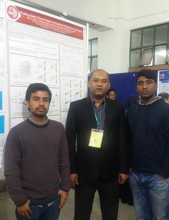
Probing Deep Tissue Organ by Electrical Impedance Technique
Abstract
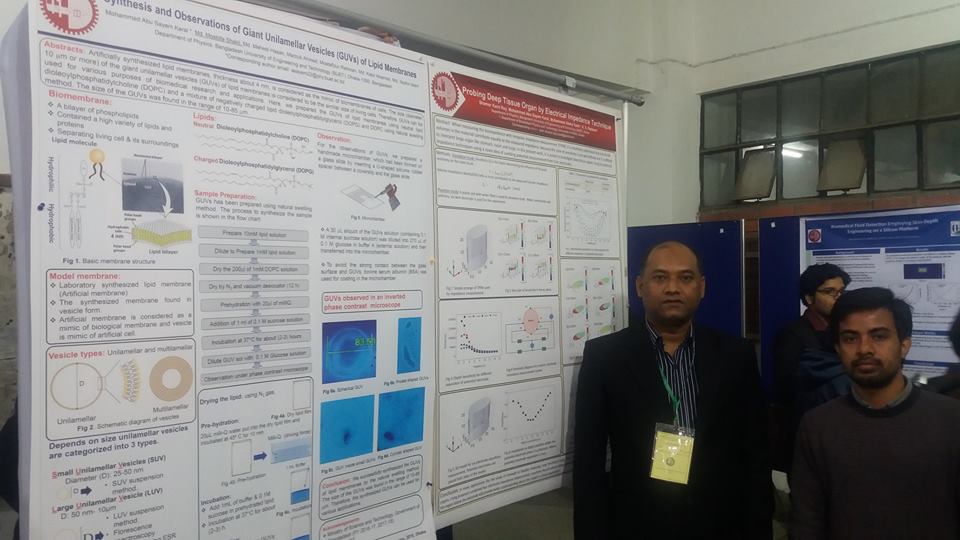
Synthesis and Observations of Giant Unilamellar Vesicles (GUVs) of Lipid Membranes
Abstract
A Facial Synthesis of Magnetite Nanoparticles Using Ipomoea aquatica Aqueous Extract and Its Anti-Bacterial Activity
Abstract
A Novel Six Electrode System for Probing Deep Tissue Organ by Electrical Impedance Technique
Abstract
A Comparative Study on Antibiotic-Induced Killing of Bacteria
Abstract
A New Six-Electrode Electrical Impedance Technique for Probing the Deep Tissue Organ of the Human Body
Abstract
Synthesis of Lipid Membranes of Giant Unilamellar Vesicles (GUVs) of and its Interactions with Magnetic Nanoparticles
Abstract
Molecular Transport through a Nano-sized Pore in the Model Membranes Using COMSOL Multiphysics
Abstract
Non-Electromechanical Technique for the Purification of Giant Unilamellar Vesicles (GUVs)
Abstract
Effects of Electrostatic Interaction on the Sizes of Giant Unilamellar Vesicles (GUVs)
Abstract
Irreversible Electroporation (IRE) Technique for the Study of Pore Formation in the Lipid Membranes of Giant Unilamellar Vesicles (GUVs)
Abstract
Edge Detection of Peptide-Induced Submicron Pores in the Lipid Membranes through ImageJ
Abstract
Elementary processes of antimicrobial peptide magainin 2-induced pore formation and its mechanism
Abstract
A biophysical approach for investigation of the antibiotic-induced bacterial killing mechanism using E. coli
Abstract
Antimicrobial peptide magainin 2-induced leakage from single E.
Abstract
Elucidation of mechanism of the effects of constant tension-induced rupture formation in giant unilamellar vesicles (GUVs) of lipid membranes using activation energy
Abstract
Effect of lipid composition on the entry of cell-penetrating peptide oligoarginine (Rn) into single vesicles
Abstract
Effect of Asymmetric Packing of Lipids in Outer and Inner Monolayer on Magainin 2-Induced Pore Formation in Lipid Bilayer
Abstract
Effect of Asymmetric Packing of Lipids in Outer and Inner Monolayer on Magainin 2-Induced Pore Formation in Lipid Bilayer
Abstract
Effects of mechanical property of lipid membranes on the entry of cell-penetrating peptide oligoarginine into a single vesicle
Abstract
Effects of Gd on Cr Doped Multiferroic BiFeO3 Ceramics
Abstract
Structural, Magnetic and Dielectric Properties of Polycrystalline La0.70Ca0.10+xSr0.20-xMnO3
Abstract
Structural and Magnetic Properties of Mn Substituted Nanocrystalline Nicuzn Ferrites
Abstract
Synthesis and Characterization of Zn Substituted Li-Ni Ferrites
Abstract
Influence of Li1+ Substitution on Impedance Spectroscopy and Electric Modulus Studies of LixCu0.10Co0.1Zn0.8-2xFe2+XO4
Abstract
Synthesis and Characterization of Bi1-xYxFe0.7Mn0.3O3 Ceramics
Abstract
Activation Energy of Tension-Induced Pore Formation in Lipid Membranes
Abstract
Investigation of Constant Tension-Induced Rupture in Lipid Membranes Using Activation Energy
Abstract
Experimental Estimation of Membrane Tension Induced by Osmotic Pressure
Abstract
Effects of lipid composition on the entry of cell-penetrating peptide oligoarginine (Rn) into single vesicles
Abstract
Effects of lipid compositions on the entry of cell-penetrating peptide oligoarginine into single vesicles
Abstract
A Mechanism of Antimicrobial Peptide, Magainin 2-Induced Pore Formation in Lipid Membranes
Abstract
Effects of lipid composition on the entry of cell-penetrating peptide oligoarginine (Rn) into single vesicles
Abstract
Analysis of Constant Tension-Induced Rupture in Lipid Membranes Using Activation Energy
Abstract
Effect of Osmotic Pressure on Constant Tension-Induced Pore Formation in Lipid Membranes
Abstract
Electrostatic Effects on Tension-Induced Pore Formation in Lipid Membranes
Abstract
Elucidation of the Mechanism of Pore formation of the Antimicrobial peptide, Magainin 2 using Single GUVs
Abstract
Electrostatic Effects on Tension-Induced Pore Formation in Lipid Membranes
Abstract
Electrostatic Effects on Tension-Induced Pore Formation in Lipid Membranes
Abstract
Effects of Line Tension on Antimicrobial Peptide Magainin 2-Induced Pore Formation
Abstract
Activation Energy of the Tension-Induced Pore Formation in Lipid Membranes
Abstract
Measurement of the Activation Energy of Tension-Induced Pore Formation in Giant Unilamellar Vesicles of Lipid Membranes
Abstract
Stretch-Activated Pore in Antimicrobial Peptide, Maganin 2
Abstract
Stretch-Activated Pore in Antimicrobial Peptide, Maganin 2
Abstract
Effects of electrostatic interactions on tension-induced pore formation in single GUVs
Abstract
Effects of electrostatic interactions on the rate constant of tension-induced pore formation in lipid membranes
Abstract
Elucidation of the Mechanism of Pore formation of the Antimicrobial peptide, Magainin 2 using Single GUVs
Abstract
Theory on the electrostatic effects on tension-induced pore formation in lipid membranes
Abstract
Stretch-Activated Pore in Antimicrobial Peptide, Maganin 2
Abstract
Effects of tension on entry of cell-penetrating peptide transportan 10 into a single vesicles and its pore formation in lipid membranes
Abstract
Effects of Mechanical Properties of Lipid Membranes on Antimicrobial Peptide Magainin 2-Induced Pore Formation
Abstract
The Single GUV method for Probing Elementary Processes of Peptide/Proteins-Induced Pore Formation in Biomembranes
Abstract
Effect of Electrostatic Interactions on Tension-Induced Pore Formation in Single GUVs
Abstract
Rate Constants of Tension-Induced Pore Formation in Lipid Membranes
Abstract
Effects of Electrostatic Interactions on Rate Constants of Tension-Induced Pore Formation in Single GUVs
Abstract
Crystallization phenomenon and magnetic properties of the amorphous (Fe(1-x)Mnx)75P15C10 alloya
Abstract
Transport properties of Fe63.75V11.25P15C10
Abstract
Characterization of amorphous Fe69V6P15C10 metallic alloys
Abstract
Structural, thermal and magnetic properties of (Fe(1-x)Mnx)75P15C10 alloys
Abstract
Magnetization, Magnetoresistance and Hall Resistivity of {Fe(100-x)Vx}75P15C10 Amorphous Ribbons
Abstract
Effect of Mn on the thermal, transport and magnetic properties of Fe0.75P0.15C0.10
Abstract
AC Properties of Mn0.5Zn0.5Fe2O4
Abstract
Transport and magnetic properties of (Fe0.70Mn0.30)0.75P0.15C0.10 alloy
Abstract
Resistivity, Magnetoresistance and Magnetization measurement of {Fe(100-x)Vx}75P15C10 Amorphous Ribbons
Abstract
Electrical, Magnetic and Thermal Properties of {Fe(1-x)Mnx}75P15C10
Abstract
Transport, magnetic and thermal properties of (Fe100-xVx)75 P15C10 alloys
Abstract
Transport and magnetic properties of double exchange (Fe1-xMnx)75P15C10 amorphous ferromagnetic alloys
Abstract
Sensitivity of the new four-electrode Focused Impedance Method (FIM) for objects with different conductivity
Abstract
Transport and magnetic properties of magnetically ordered (Fe100-xVx)75P15C10 amorphous alloys
Abstract
Structural, dielectric and electrical properties of Zn1-x-yCdxLiyO system
Abstract
Conductivity dependent sensitivity in 4-electrode Focused Impedance Measurement (FIM) system
Abstract
Magnetic and electrical properties of Fe76.5-xNbxSi13.5B9Ag1 alloys
Abstract
A new four-electrode Focused Impedance Measurement (FIM) system for physiological study
Abstract
Abstract
বিশ্ববিদ্যালয় ও কলেজ পর্যায়ের স্নাতক ও স্নাতকোত্তর শ্রেণির ফিজিক্স, মেডিকেল ফিজিক্স, বায়োমেডিকেল ফিজিক্স ও বায়োমেডিকেল ইঞ্জিনিয়ারিং বিভাগের সিলেবাস অনুসারে বইটি লেখা। এতে ষোলোটি অধ্যায়। প্রথম আট অধ্যায়ে বায়োফিজিক্স নিয়ে আলোচনা রয়েছে। শেষ আট অধ্যায়ে আলোচনা করা হয়েছে মেডিকেল ফিজিক্স নিয়ে। বায়োফিজিক্স অংশে ম্যাক্রোমলিকিউলস, নিউক্লিক অ্যাসিড, প্রোটিনের গঠন, এনজাইমের মৌলিক আচরণ, কোষীয় মেমব্রেন, নার্ভাস সিস্টেম, মাসল এবং কার্ডিও-ভাসকুলার সিস্টেম সম্পর্কে বর্ণনা রয়েছে। আর মেডিকেল ফিজিক্স অংশে আলট্রাসাউন্ড ইমেজিং, অন্যান্য ইমেজিং কৌশল, ওডিওলজি, ভাসকুলার পরিমাপ, কার্ডিয়াক পরিমাপ, নিউরোমাসকুলার পরিমাপ, বায়োইলেকট্রিক অ্যামপ্লিফায়ার এবং রেডিয়েশন ও স্বাস্থ্য নিয়ে আলোচনা আছে। প্রয়োজনীয় চিত্র এবং গাণিতিক সমীকরণ বইটির গুরুত্ব বাড়িয়েছে।
Biomedical Physics has been published from a country renowned publishing house ‘Prothoma Prokashan’. This book will be helpful for the students and teachers in all public and private universities and colleges in the undergraduate and graduate level. This is the first book written in Bangla in this field. The book is available in Prothoma Prokashan, Aziz Super Market, Shahbag, Dhaka; and Prothom Alo office at Karwan Bazar, Dhaka, Bangladesh
Recent Posts
Research Summary
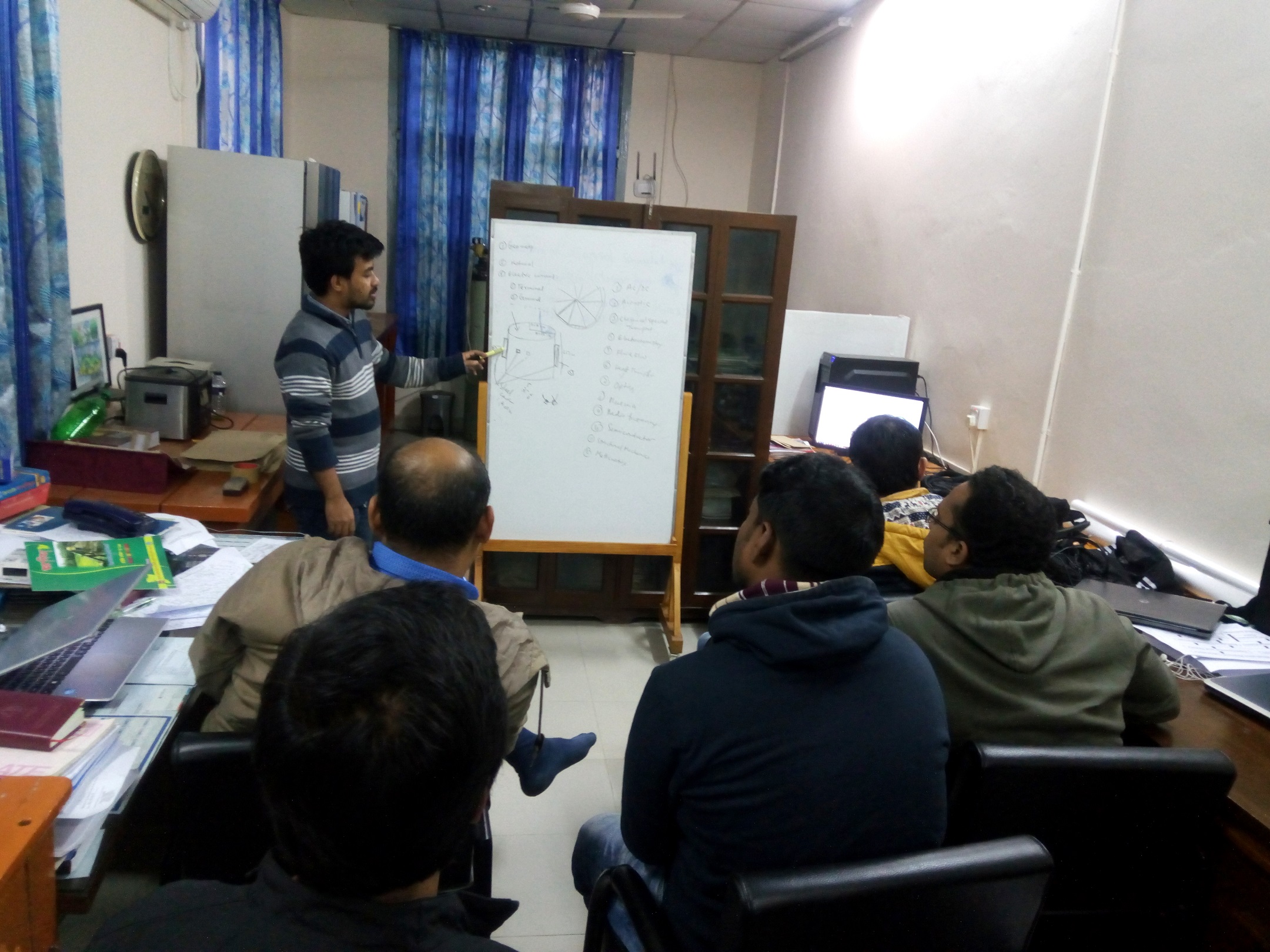
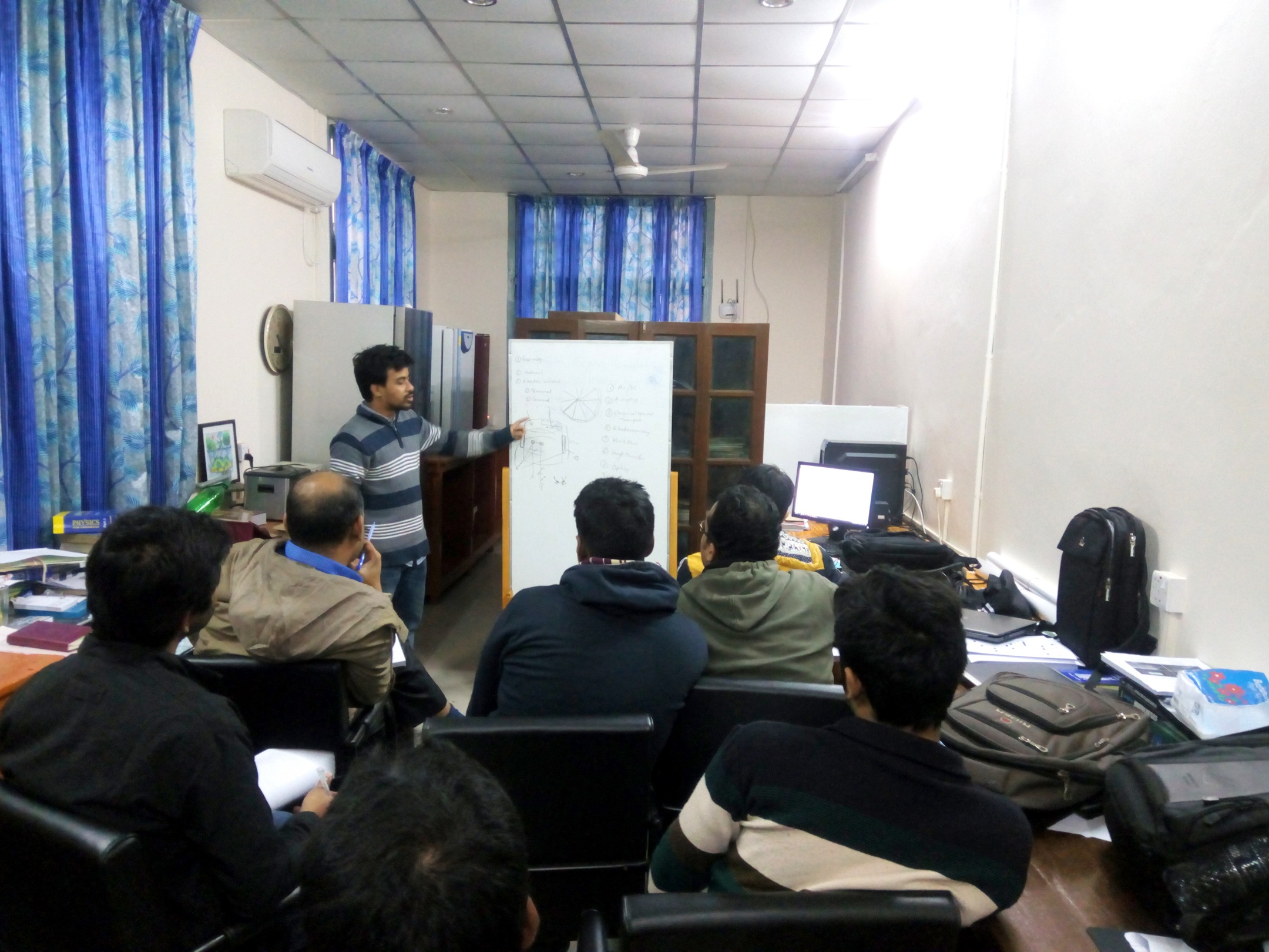
The Biophysics Research Laboratory of the Department of Physics of BUET is one of the leading research Lab in Bangladesh. The Laboratory is keen on pushing the boundaries of traditional research system and vigilant to face the challenges that the ever changing field of research brings forth. It is the optimum combination of knowledge generation and application that makes the distinctive feature of the Physics Department in BUET.
Interests
- Membrane Biophysics
- Nanotechnology
- Medical Physics
- Biomedical Physics
- Health Physics
- Nuclear Physics
- Simulation
Laboratory Personel
Biophysics Research Laboratory
The Laboratory focuses heavily on the quality and impact of the researches done in the field of Biophysics as well as in multidisciplinary researches
+ FollowBiophysics Research Laboratory mainly deals with artificial lipid membranes synthesis and its characterization. The lipid membranes has been prepared in the form of vesicles. It is also called liposome. Vesicles or liposomes can be categorized into three types depending on their size. It would be giant unilamellar vesicles (GUVs), large unilamellar vesicles (LUVs) and small unilamellar vesicles (SUVs). The size of the GUVs is very similar to real cell and the physiological condition of GUVs is also similar to cell. Therefore, GUVs can be used a artificial cells and the lipid membranes is considered as a mimic of plasma membranes or biomembranes of cells. We also deals with nanoparticles synthesis, characterization and its biological applications
Research Projects
-
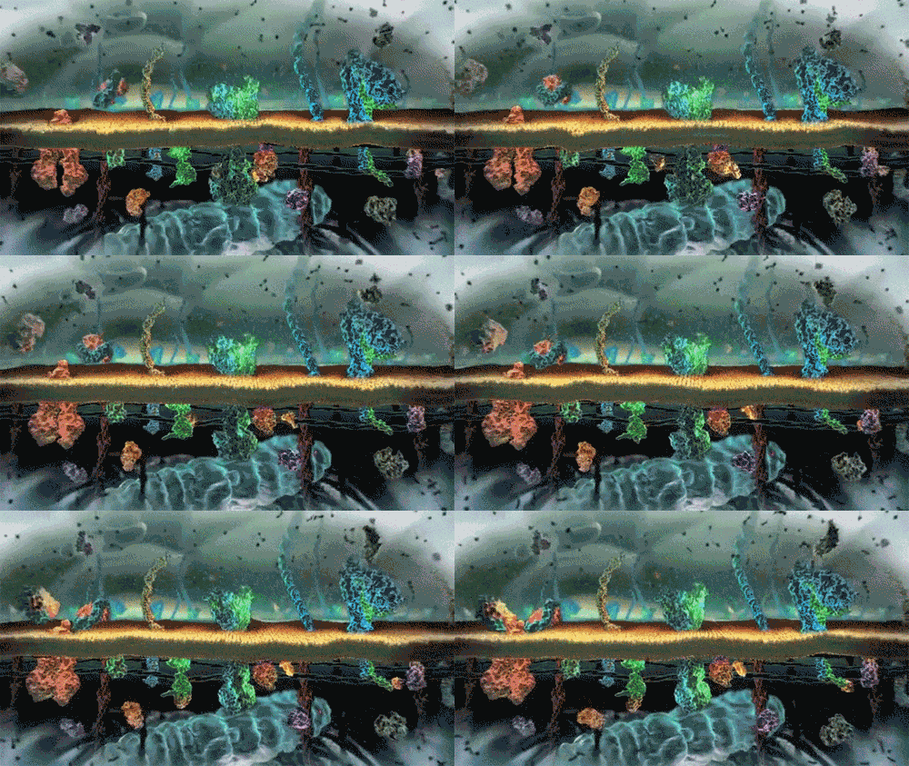
Biophysics or biological physics is an interdisciplinary science that applies the approaches and methods of physics to study biological systems. Biophysicists study life at every level, from atoms and molecules to cells, organisms, and environments. As innovations come out of physics and biology labs, biophysicists find new areas to explore where they can apply their expertise, create new tools, and learn new things. The work always aims to find out how biological systems work.
-
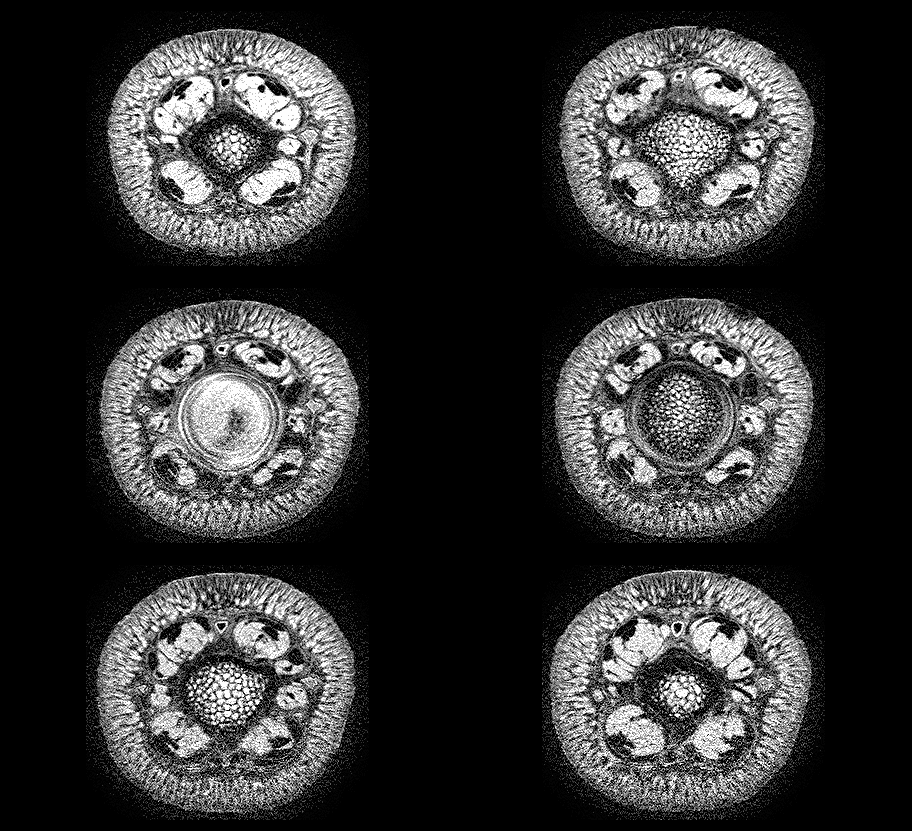
The biomedical physics seeks to understand the role of physical processes occurring on molecular, cellular, or macroscopic scales; ranging from the interaction of chemicals with DNA, to the generation of complex electrical signals in the brain and nervous system; and physical principles of medical devices. It also includes the innovations of hardware and software development whether it can be used for future clinical applications or research in animals and humans.
-
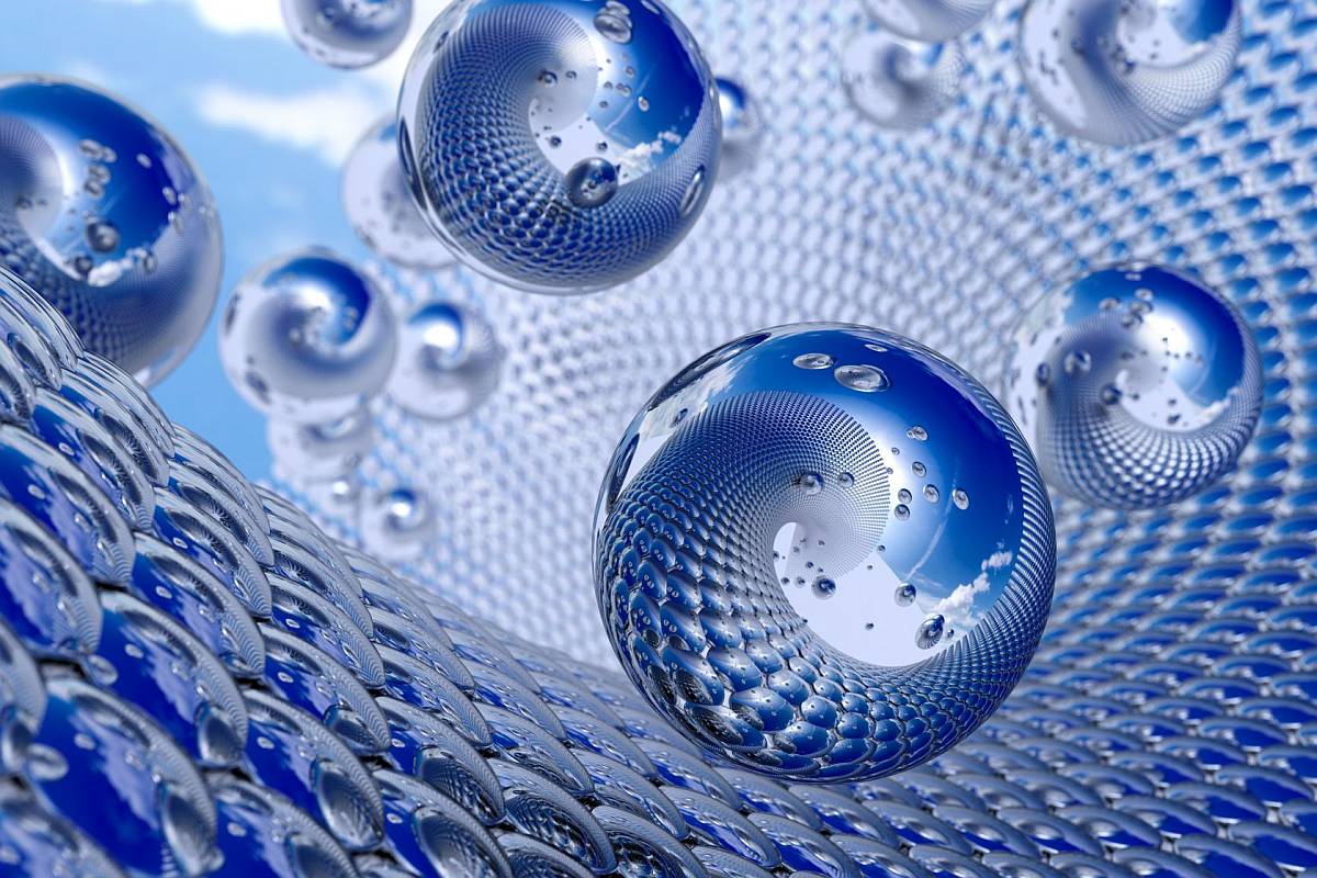
Nanobiomaterials exhibit distinctive characteristics, including mechanical, electrical, and optical properties, which make them suitable for a variety of biological applications. Because of their versatility, they play a central role in nanobiotechnology and make significant contributions to biomedical research and healthcare. Novel approaches for bottom-up and top-down processing of nanostructured biomaterials are highlighted.
-
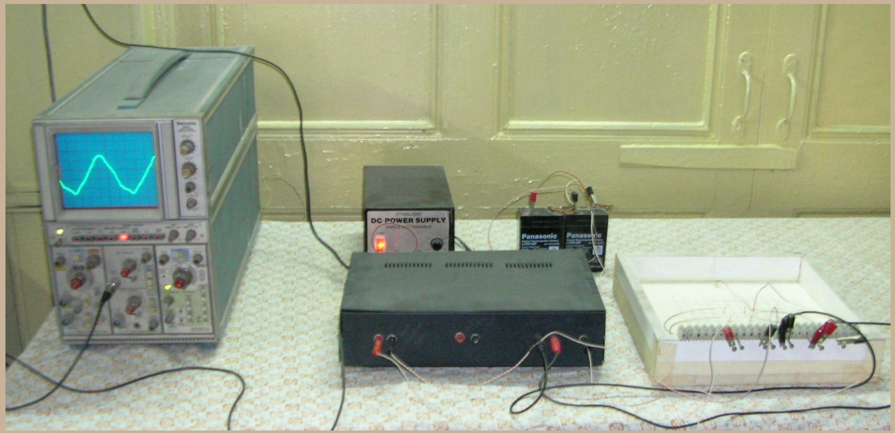
Bioinstrumentation is the use of bioelectronic instruments for the recording or transmission of physiological information. Biomedical devices are an amalgamation of biology, sensors, interface electronics, microcontrollers, and computer programming, and require the combination of several traditional disciplines including biology, optics, mechanics, mathematics, electronics, chemistry, and computer science. Bioinstrumentation teams gather engineers that design, fabricate, test, and manufacture advanced medical instruments and implantabe devices into a single, more productive unit.
-
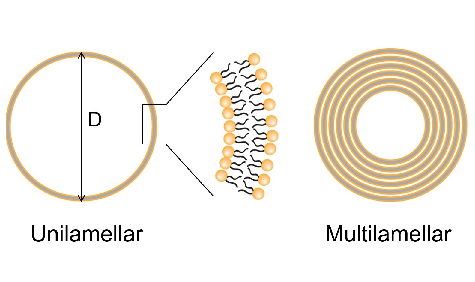
Artificially synthesized lipid membranes of thickness about 4 nm are considered as the mimic of biomembranes of cells. The size (diameter 10 micrometer or more) of the giant unilamellar vesicles (GUVs) of lipid membranes is considered to be the similar size of living cells. Therefore, GUVs can be used for various purposes of biomedical research and applications. Here, we prepared the GUVs of lipid membranes using neutral lipid dioleoylphosphatidylcholine (DOPC) and a mixture of negatively charged lipid dioleoylphosphatidylglycerol (DOPG) and DOPC using natural swelling method. The size of the GUVs was found in the range of 10-85 micrometer

